← anatomy of the ear mcat Dissect a human ear « science experiments :: wonderhowto Lobe Lobes brain frontal temporal lobe occipital parietal four green cortex cerebral anatomy red cognition include yellow functions motor movement right →
If you are searching about Ear Diagram Round Window - Human Anatomy you've visit to the right place. We have 35 Pictures about Ear Diagram Round Window - Human Anatomy like | Illustration of the inner ear, with highlights of the oval (OW) and, Surgery | Hyperacusis Focus and also Middle ear - Wikipedia. Here you go:
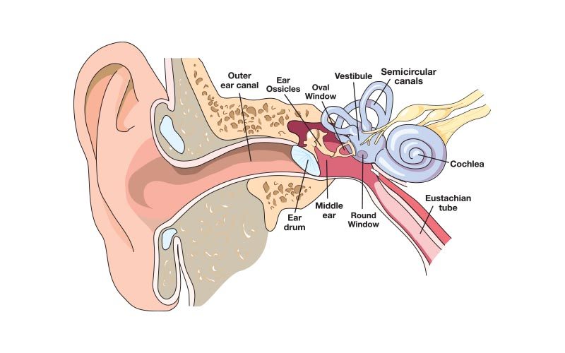
Ear Diagram Round Window - Human Anatomy
 tartrerepub.blogspot.com
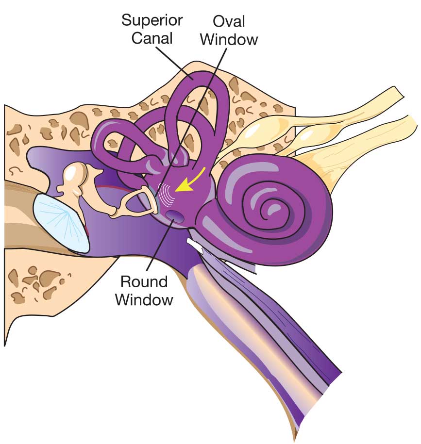
Prominence Of Lateral Semicircular Canal Superior To Oval Window
 www.pinterest.com
oval windows ear fenestra middle window round vestibuli cochlea promontory located medical inner tympanic membrane hidden bones shaped turn scala
Sweetibnotes: Option E - Neurobiology And Behaviour
 sweetibnotes.blogspot.ca
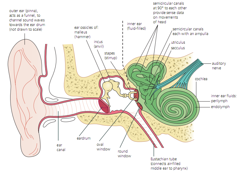
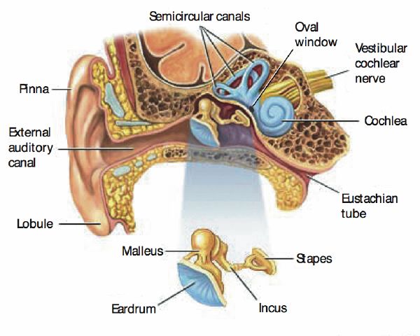
ear window oval round diagram label cochlea middle pinna eardrum canals semicircular
Physiology Of Hearing
 www.onlinebiologynotes.com
hearing physiology receptors ear structure cochlea perilymph auditory membrane location uncoiled sound anatomy vibration sensory tympanic involved internal external pressure
Anatomic Structure Of The Human Ear. Diagram Of The Ear And Organ Of
 www.researchgate.net
ear corti oval anatomic anatomy retrieved slidedocnow stapes
Middle Ear - Wikipedia
 en.wikipedia.org
ear middle anatomy human
Hearing And Vestibular Sensation | Biology II
 courses.lumenlearning.com
vestibular ear biology sound window round cochlea human membrane parts tympanum through sensation part called hearing figure pinna nerve canal
Hearing | Introduction To Psychology
 courses.lumenlearning.com
ear membrane ossicles tympanic inner incus stapes malleus pinna auditory cochlea basilar psychology middle into sound window oval waves labeled
Lab Guide: Ear, Eye, Brain
 www.biologycorner.com
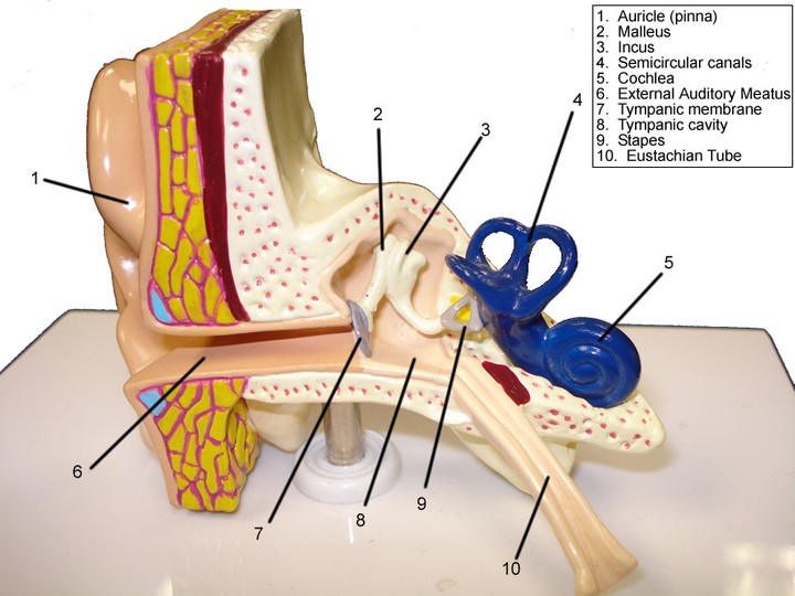
ear anatomy eye lab models guide model biologycorner structures parts identify senses brain physiology eyes biology use test answers specimens
PPT - Bio 449 Lecture 11 - Sensory Physiology III Sep. 20, 2010
 www.slideserve.com
ear anatomy physiology sensory ppt inner lecture bio iii sep 2010 gustation bone middle cochlea powerpoint presentation external slideserve
How You Hear - Northland Audiology
 northlandaudiology.com
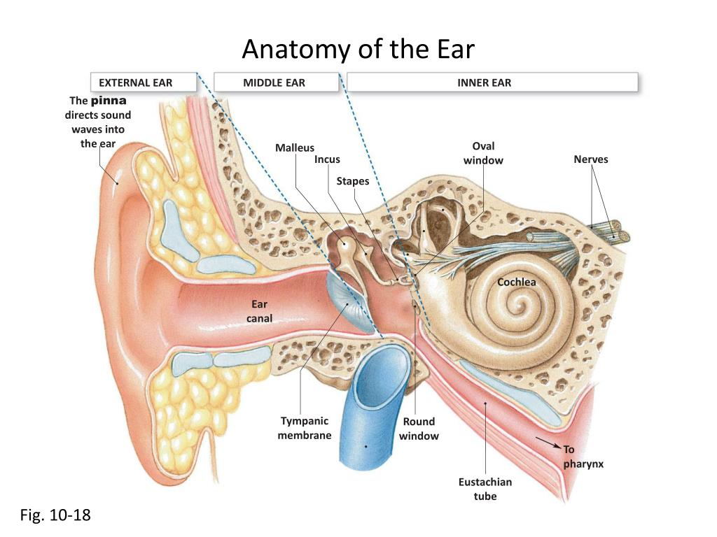
ear diagram hear function inner canal anatomy labels hearing chart loss northland flow assignment
Ghim Trên Anatomy/medicine
 www.pinterest.com
ear anatomy endolymph perilymph membranous round uni doctors window oval csf cochlear labyrinth vestibular vestibule diagram semicircular its blogger stapedial
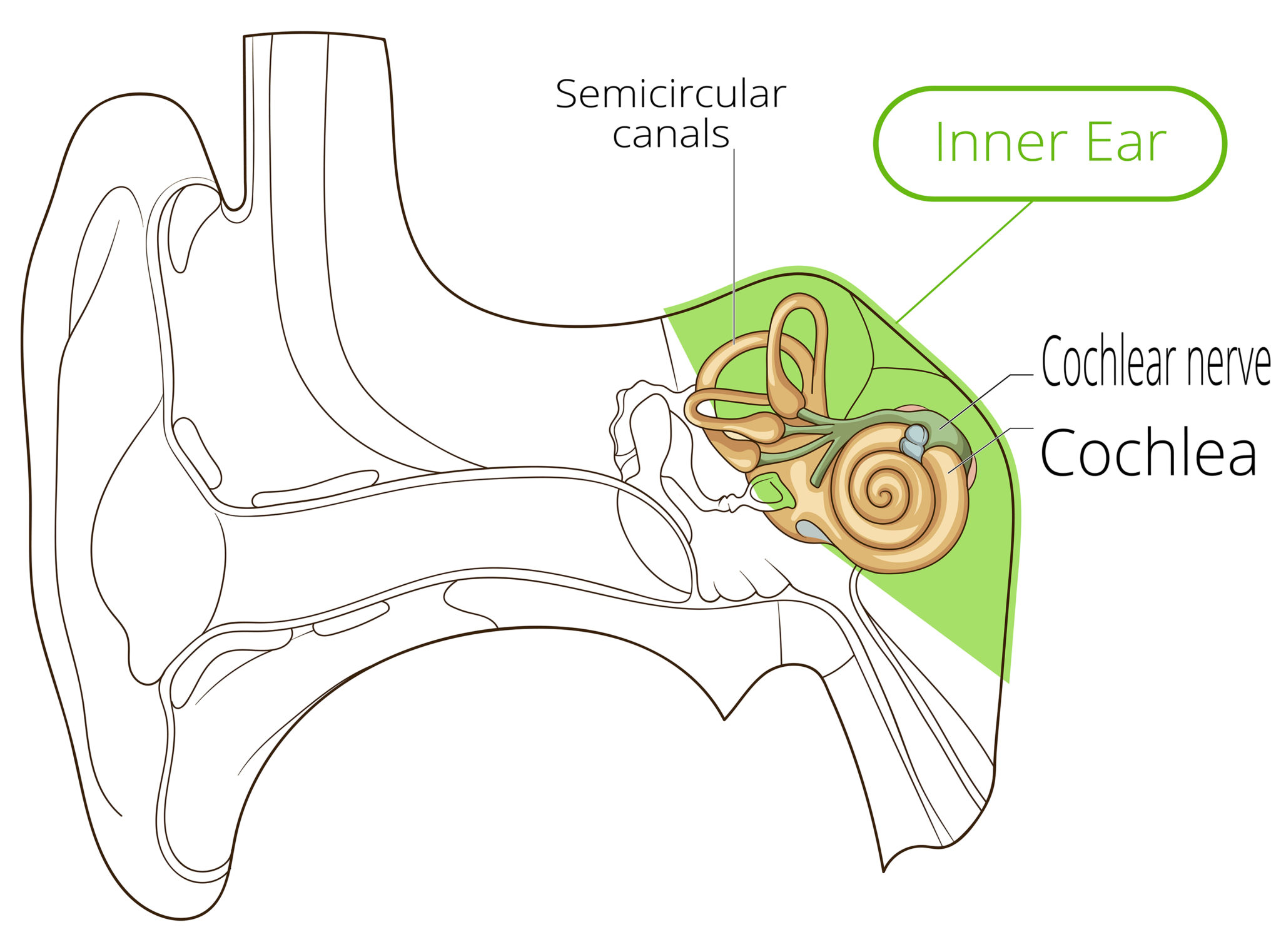
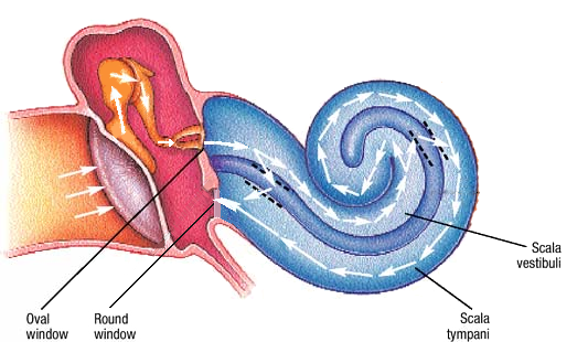
| Illustration Of The Inner Ear, With Highlights Of The Oval (OW) And
 www.researchgate.net
ow cochlear perilymph endolymph
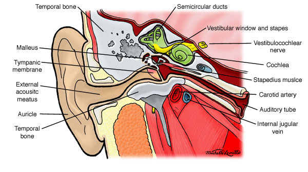
Ear Anatomy | Causes Of Hearing Loss | Hearing Aids | Audiology
 www.lakeenthearing.com
telinga inner fungsinya middle gooddoctor anatomi canals pengertian meliputi labirin semicircular
Ear Diagram Ap Psych - Human Body Anatomy
 gentleforindustrial.blogspot.com
psych vestibular stapes
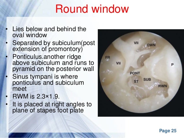
Round Window | Anatomy | Britannica
 www.britannica.com
britannica membrane auditory cochlea corti basilar ossicles nerve
Attic Area Of Tympanic Membrane - Image Balcony And Attic
 aannemerdenhaag.org
tympanic attic physiology auditory ento
Ear Anatomy
 www.edoctoronline.com
ear anatomy middle window round oval inner stapes auditory ossicles cochlea malleus vestibule incus canals semicircular hearing human atlas f4
Enhanced Viral-mediated Cochlear Gene Delivery In Adult Mice By
 www.nature.com
mice window round canal membrane fenestration inoculation cochlear adult gene
Ear Diagram Oval And Round Windows - Diagram Media
 diagramedia.blogspot.com
illustrating therapeutics
Inner Ear Diagram Oval Window - Diagram Media
 diagramedia.blogspot.com
Inner-Ear Barotrauma (IEBT) | Ears & Diving - DAN Health Issues & Diving
 world.dan.org
Anatomy Of The Human Round Window (left Ear-medial View). A, The RW Is
medial
First Aids - Catalao.cml
 sites.google.com
ear external anatomy base study aid approach peer
Endoscopic View Of The Right Middle Ear. S; Stapes, P: Promontorium; R
 www.researchgate.net
stapes endoscopic promontorium tendon
Surgery | Hyperacusis Focus
 hyperacusisfocus.org
reinforcement surgical
Perilymph Fistula Causes, Symptoms, Diagnosis, Treatment & Prognosis
 healthjade.net
fistula perilymph perilymphatic causes slidedocnow vertigo diagnosis
Medi Photos: Surface Anatomy Of The External Ear
 mediphotos.blogspot.com
ear external anatomy surface helix medi ears auricle auditory diagram rim 3d canal cgarena consists
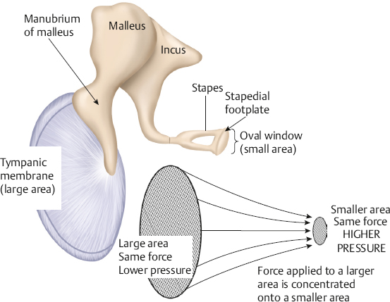
Physiology Physics Woven Fine: Hearing Involves Sound Physics
 physiology-physics.blogspot.com
ear anatomy sound hearing physics ears ossicles does window oval round physiology auditory tmj ringing cs showing involves gif hear
Tristan Jehan PhD Thesis - Chapter 3
 web.media.mit.edu
ear anatomy middle figure tristan phd listening music psychoacoustics dissertation mit edu web
Diagram Of The Inner Ear
 www.osha.gov
ear inner diagram anatomy osha balance gif sixth sense cochlea vestibular system column spirals turns bony resembles snail shell within
Round Window Niche. Right Ear: Details Of Important Middle Ear
 www.researchgate.net
niche cochlear
Ear Diagram Oval Window - Diagram Media
 diagramedia.blogspot.com
anatomia korvan barotrauma common kansalliset
Middle Ear Anatomy
 www.slideshare.net
How Your Ears Respond To Pressure - Divers Alert Network
 dan.org
equalization barotrauma equalizing divers diver squeeze equalize
Fistula perilymph perilymphatic causes slidedocnow vertigo diagnosis. | illustration of the inner ear, with highlights of the oval (ow) and. Sweetibnotes: option e