← anatomy of the eyeball Anatomy eyeball gross anatomy of the eye diagram Eye diagram human anatomy eyes camera layer vision →
If you are looking for Normal MR Imaging Anatomy of the Knee - Magnetic Resonance Imaging Clinics you've visit to the right web. We have 35 Images about Normal MR Imaging Anatomy of the Knee - Magnetic Resonance Imaging Clinics like Figure. Anatomy of the Right Knee | Download Scientific Diagram, Knee Pain: Proximal Tibiofibular Subluxation - Life Hypermobile and also Back Bones : In this article, we explain their function, what they are. Here you go:
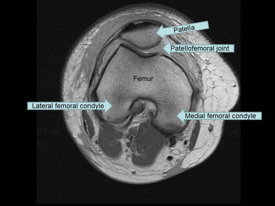
Normal MR Imaging Anatomy Of The Knee - Magnetic Resonance Imaging Clinics
 www.mri.theclinics.com
knee mri anatomy axial patellofemoral normal cartilage femoral
Figure. Anatomy Of The Right Knee | Download Scientific Diagram
 www.researchgate.net
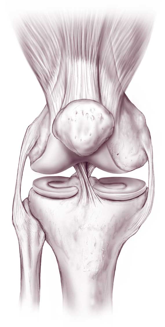
meniscus tendon patellar ligament structures
1000+ Images About Anatomy & Radiology On Pinterest | Bursa, Pain D
 www.pinterest.com
knee anatomy sagittal mri cross sectional radiology mrimaster section ct side use tool mouse study choose board move
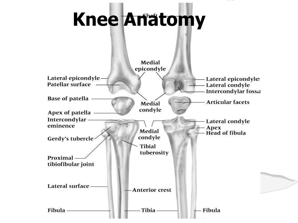
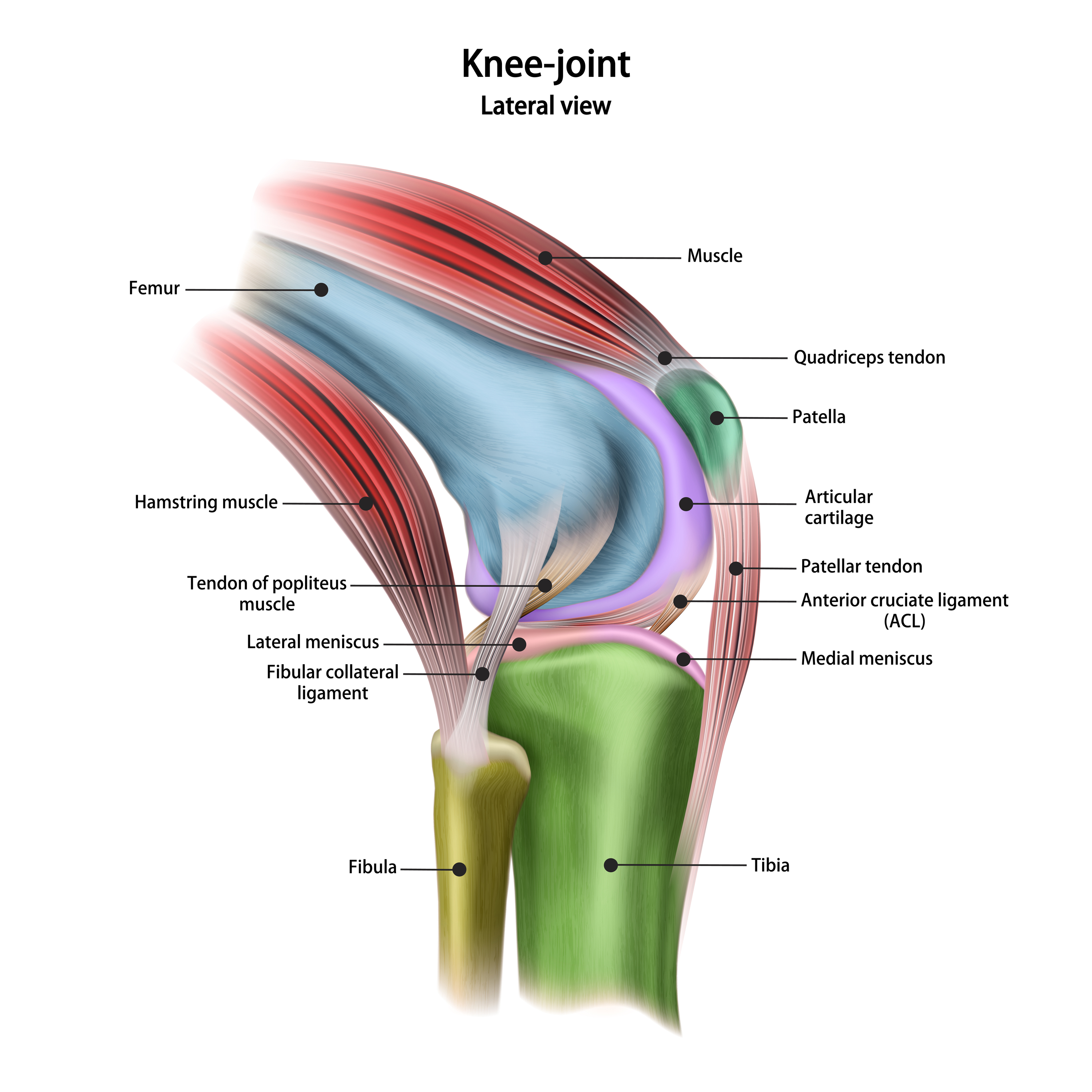
Knee Anatomy
 www.joionline.net
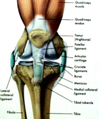
knee anatomy ligaments muscles joint bones labels tendons cartilage joints injury
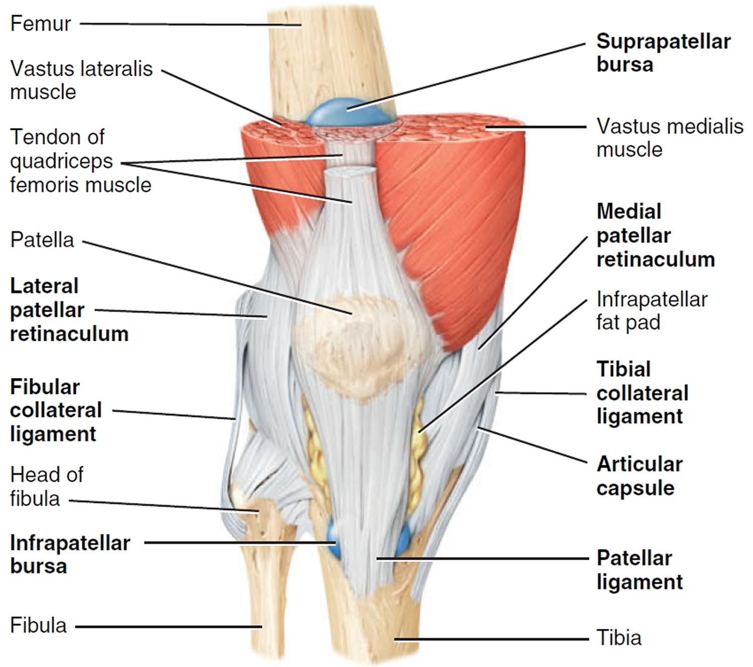
Knee Anatomy: Passive Structures - Bones, Ligaments, Meniscus
 www.fix-knee-pain.com
knee anatomy structures ligaments passive schematic ligament human pain lateral collateral joint diagram cartilage anterior body anatomical leg bones part
Knee Pain - Causes, Exercises, Remedies, Medication & Treatment
 healthjade.net
knee anatomy posterior joint pain figure sagittal
Back Bones : In This Article, We Explain Their Function, What They Are
 kylerjourney.blogspot.com
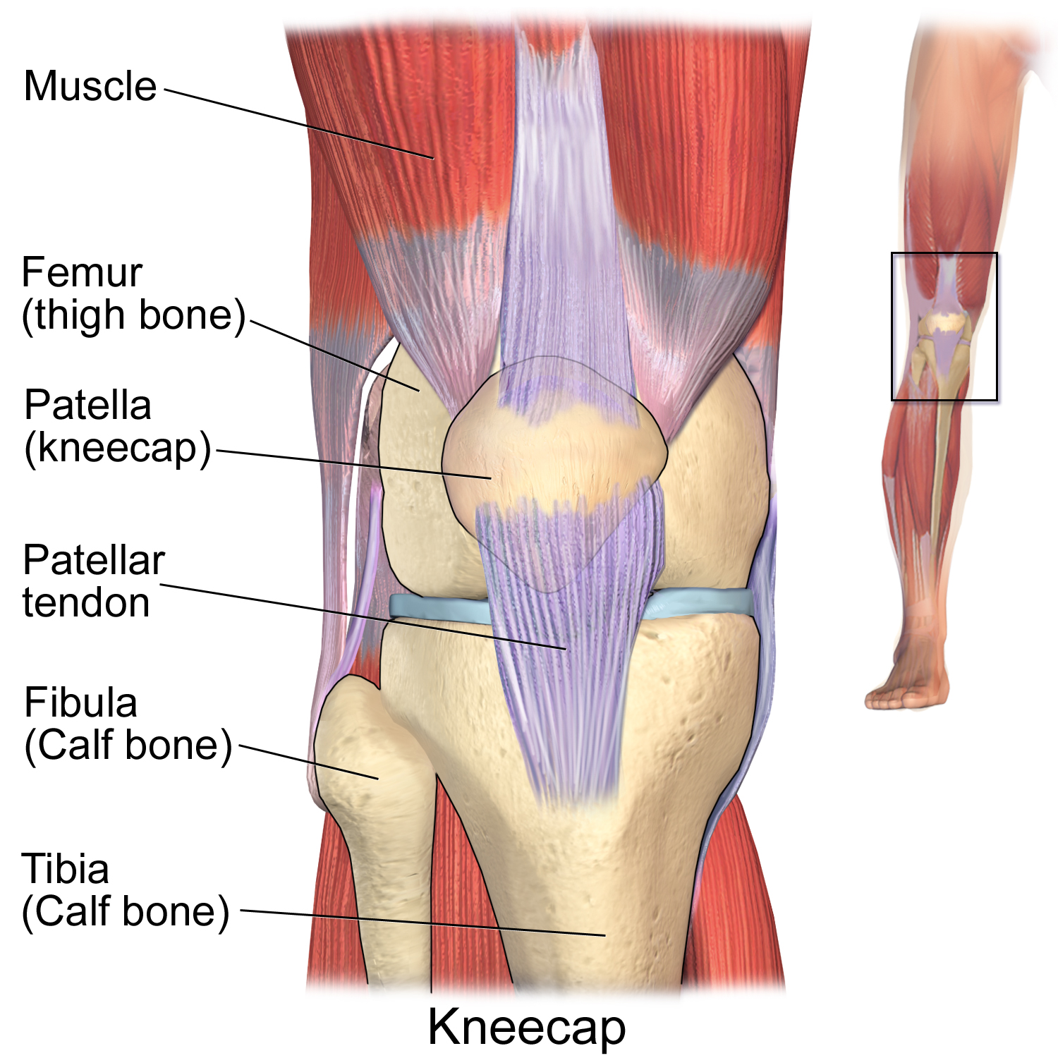
bone patella upper tendon lateral orthopedic femur gachou22 tendons
What Is Causing Your Knee Pain?
:max_bytes(150000):strip_icc()/188058334-crop-56aae7425f9b58b7d0091480.jpg) arthritis.about.com
arthritis.about.com
pain stocktrek
Magnetic Resonance Imaging - Knee Injury And Prevention
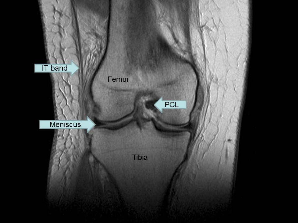 kneeinjury.weebly.com
mri coronal resonance menisci sagittal
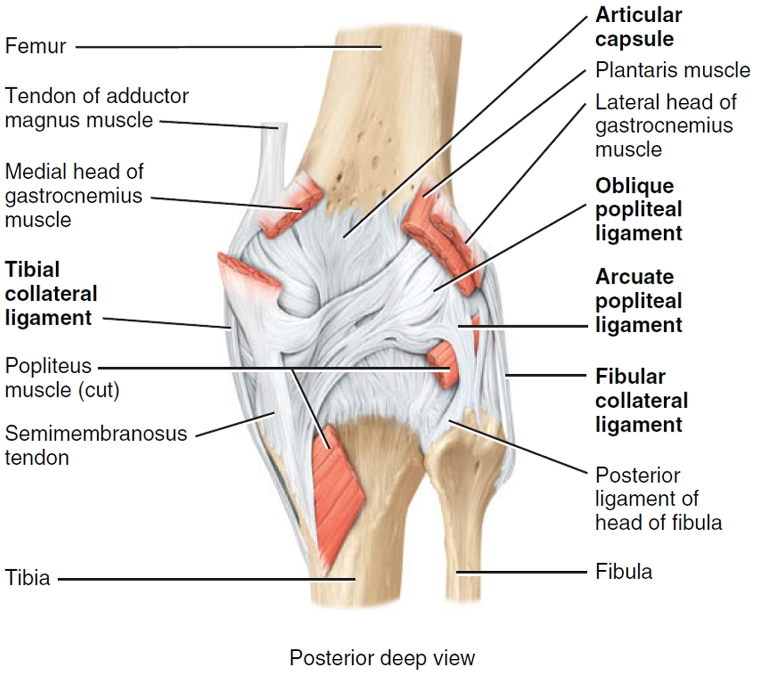
Right Knee, Posterior View
 www.humangrossanatomy.us
posterior extremity ligament medial netter
Anatomy Of The Right Knee - TrialExhibits Inc.
 www.trialexhibitsinc.com
Anatomy – Page 65 – Graph Diagram
 graphdiagram.com
anatomy diagram knee chart muscles
On The Bench :( | Blue Runnings
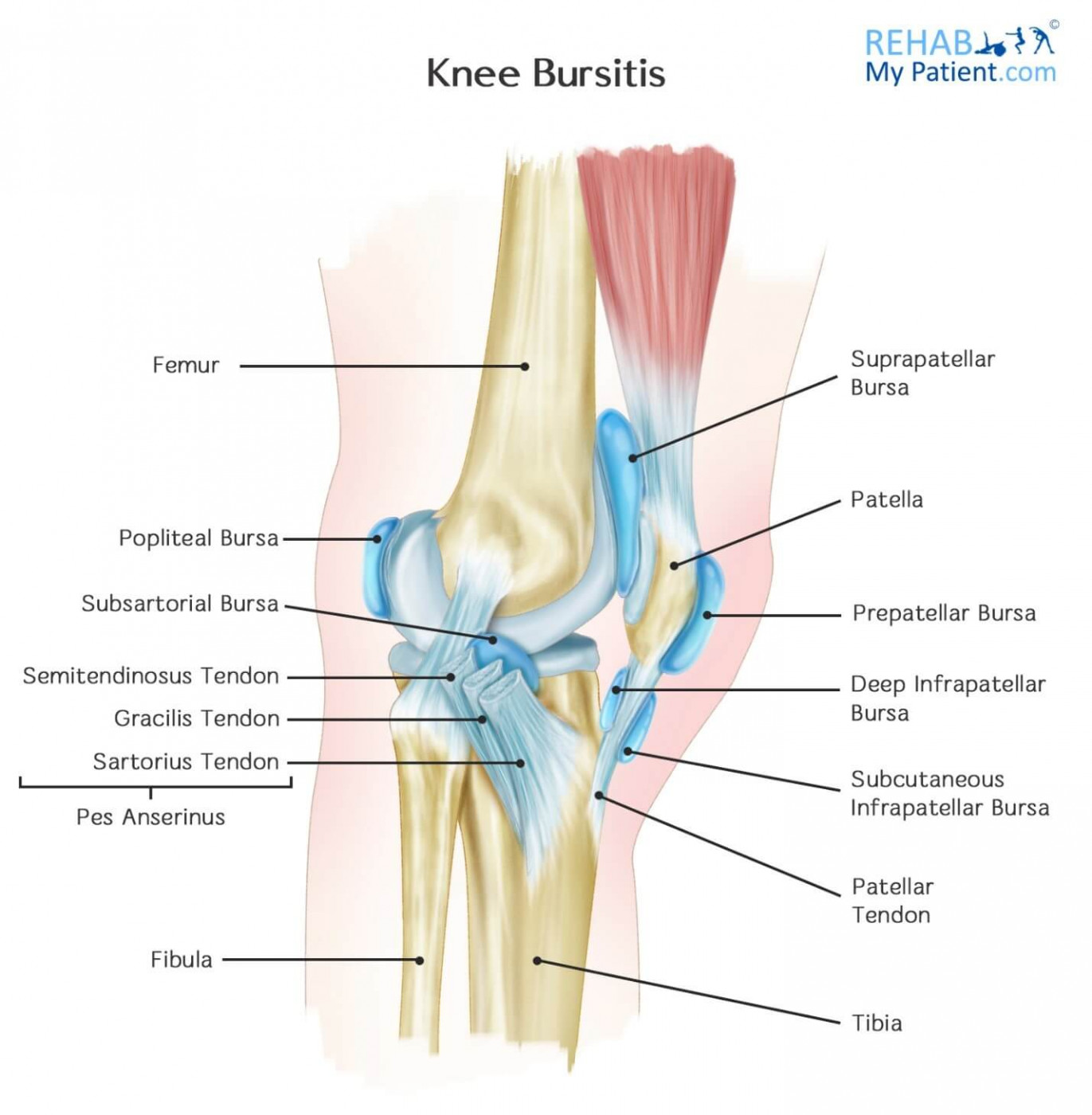 bluerunnings.com
knee bursitis bursa fluid kneecap articles runnings blue
Knee Arthroscopy | Pictures, Recovery, Ligament, Meniscus, Chondromalacia
 www.healthpages.org
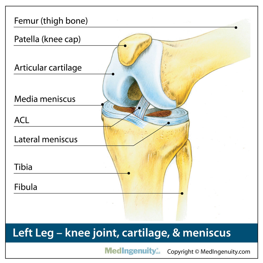
arthroscopy anatomie knies meniscus ligament healthy
PPT - Knee Anatomy PowerPoint Presentation, Free Download - ID:5326168
 www.slideserve.com
knee anatomy femur bones tibia joint ppt distal fibula anatomie patella presentation flat fractures powerpoint femoral base poorly constructed most
Normal MR Imaging Anatomy Of The Knee - Magnetic Resonance Imaging Clinics
 www.mri.theclinics.com
knee mri anatomy axial patellofemoral muscle normal compartment fig ppt imaging figure viewer hi res mr
Anatomy Library | Fort Worth Bone & Joint Clinic
 thcboneandjoint.com
anatomy knee joint library bone cartilage replacement
Knee Pain: Proximal Tibiofibular Subluxation - Life Hypermobile
 lifehypermobile.com
knee pain proximal tibiofibular anatomy diagram subluxation basic
Anatomy Of The Knee Posterior View - Life Educations
 thelifeedu.blogspot.com
knee posterior limb
Knee Anatomy - Posterior View - TrialExhibits Inc.
 www.trialexhibitsinc.com
8 Lateral Anatomy Of The Right Knee Of A Human Cadaver In A Extension
 www.researchgate.net
knee lateral popliteus cadaver posteromedial anterolateral inserted arthroscope distal flexion
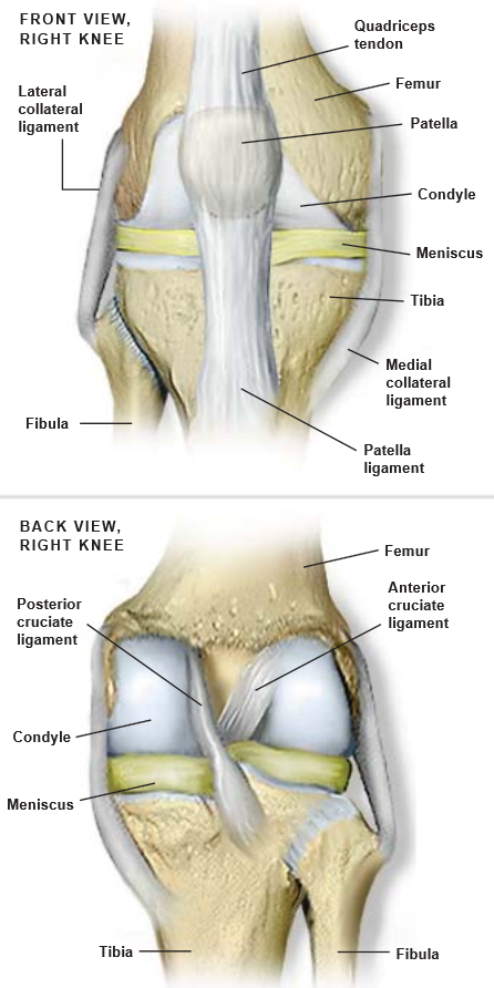
Front View Of Knee Anatomy [11] | Download Scientific Diagram
![Front view of knee anatomy [11] | Download Scientific Diagram](https://www.researchgate.net/profile/Jonisha_Pollard/publication/262301586/figure/download/fig4/AS:300870114791426@1448744433151/Front-view-of-knee-anatomy-11.png) www.researchgate.net
www.researchgate.net
Magnetic Resonance Imaging - Knee Injury And Prevention
 kneeinjury.weebly.com
mri knee axial acl pcl joint normal interpret location muscles ligaments imaging cruciate patellofemoral comments injury example iliotibial band collateral
Knee & Leg - Atlas Of Anatomy
 doctorlib.info
anatomy knee joint leg atlas ligaments ligament right plane muscles menisci muscle tibial human tendon extension doctorlib info anatomi capsule
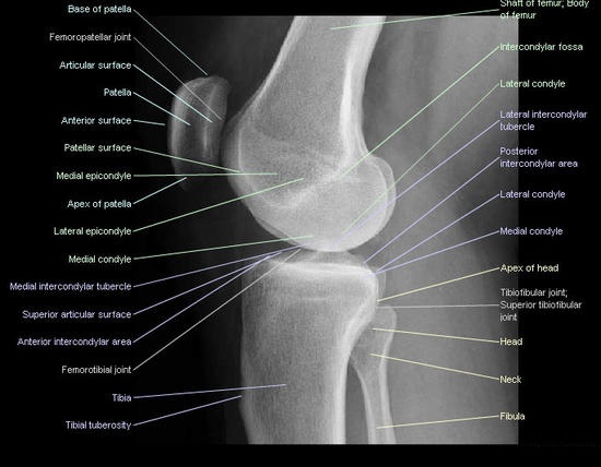
Radiographic Positioning Examples Of The Leg And Knee - CE4RT
 ce4rt.com
knee lateral anatomy lower bones ray positioning xray joint radiograph atlas limb radiographic arteries medical extremity views rays radiology leg
Knee Injuries Causes, Types, Symptoms, Knee Injuries Prevention & Treatment
 healthjade.com
knee anatomy joint injuries figure
Promatx Health Club: How Can I Strengthen My Knees?
 promatx.blogspot.com
knee anatomy joint leg palpation human knees muscle bones strengthen pain yoga bone forward structure
Science & Medicine: Knee-joint Ligaments
 science-naturalphenomena.blogspot.com
ligaments ligament tendons tendon cruciate
Pin By Cerridwen MacLeod On Radiology | Radiology Student, Medical
 www.pinterest.com
radiology knee anatomy ap student medical human physiology radiographic body
Knee Anatomy | Complete Orthopedics | Multiple NY Locations
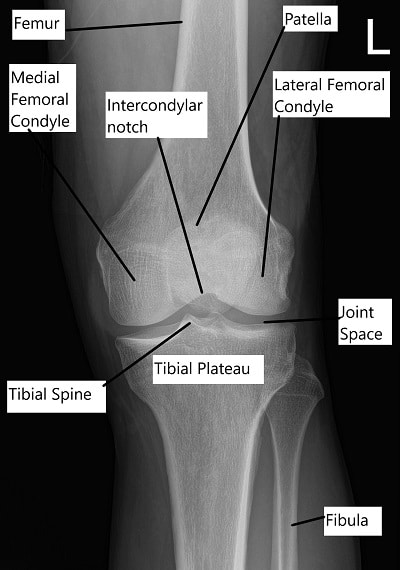 www.cortho.org
anatomy ray tibia
Does Physiotherapy Help Knee Pain? - BMJ Physiotherapy Clinic
 bmjtherapy.com
anatomy ligaments ligament hoek struktur knie knees tendons patellaspitzensyndrom injections cell musculoskeletal tendinitis joionline physiotherapy menschlichen seitenansicht medizinisch kniegelenks aller
Knee Tendon Diagram - Collateral Ligament Injuries - OrthoInfo - AAOS
 anielafarley.blogspot.com
ligament tendon tendons geometrical graft collateral aaos orthoinfo
Ultrasound Of The Knee
 www.moleopedia.com
lcl ultrasound sprain
Knee Anatomy | MRI Knee Coronal Anatomy | Free Cross Sectional Anatomy
 mrimaster.com
mri knee anatomy coronal cross sectional use move mrimaster alternatively arrows tiny both side down
Magnetic Resonance Imaging - Knee Injury And Prevention
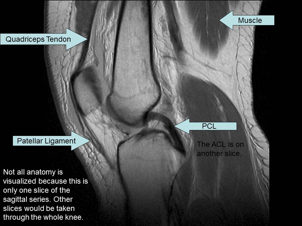 kneeinjury.weebly.com
knee mri sagittal normal acl pcl muscles ligaments injury resonance magnetic quadriceps tendon ligament imaging example patellar posterior cruciate
Mri knee axial acl pcl joint normal interpret location muscles ligaments imaging cruciate patellofemoral comments injury example iliotibial band collateral. Anatomy ray tibia. Radiology knee anatomy ap student medical human physiology radiographic body